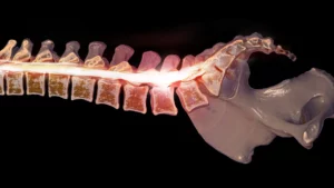
What is a Pedicle Fracture?
A fracture of pedicle, which is a bony bridge connecting the vertebral body to the posterior elements of the spine, can occur due to various stress factors or traumatic events. The pedicles are essential for the stability and structural integrity of the spine.
Pedicle fractures are relatively rare and can result from high-impact trauma, repetitive stress, or conditions that weaken the bone, such as osteoporosis. These fractures can occur in any part of the spine but are most commonly seen in the lumbar region (lower back).¹⁻³
Fracture of Pedicle Causes
Trauma
Severe trauma, such as car accidents, falls, or sports injuries, can lead to spine pedicle fractures. These incidents often result in high-impact forces that can break the pedicles, which are the bony projections on the vertebrae.²⁻⁴
Repetitive Stress
Repetitive mechanical stress, particularly in athletes who engage in activities involving hyperextension of the spine, can lead to pedicle stress fractures. This repetitive strain can cause micro-damage over time, eventually resulting in a fracture.⁴
Bone Health Issues
Conditions like osteoporosis, which weaken the bones, increase the risk of fractures even with minor trauma or stress. Osteoporosis reduces bone density, making the pedicles more susceptible to fractures. Other bone health issues, such as osteopenia and degenerative spinal diseases, can also contribute to the likelihood of fractures.¹⁻²
Abnormal Spinal Mechanics
Structural abnormalities in the spine, such as scoliosis (abnormal curvature of the spine) or improper alignment, can cause uneven stress distribution on the pedicles. This uneven stress can lead to fractures over time as the bones endure abnormal forces.¹
Post-Surgical Complications
Previous spinal surgeries, especially those involving instrumentation or fusion, can alter the stress dynamics on the spine. This can lead to adjacent segment disease, where the areas next to the fused segments experience increased stress, potentially causing pedicle fractures.²
Fracture of Pedicle Symptoms
A pedicle fracture can present with various symptoms, largely depending on the severity and specific location of the fracture. Common symptoms include:
Severe Back Pain
This is typically localized to the area of the fracture and may worsen with movement.⁵⁻⁶
Tenderness and Discomfort
The affected area may be tender to the touch, and discomfort can be persistent.¹⁻⁶
Limited Mobility
Movement, especially bending or twisting, can be significantly painful and restricted.¹⁻⁵
Neurological Symptoms
If the fracture impacts the spinal cord or nearby nerves, symptoms might include numbness, tingling, or weakness in the limbs. More severe cases can lead to bowel or bladder dysfunction.⁵⁻⁶
Muscle Spasms
Spasms or involuntary contractions of the muscles around the fracture site can occur.¹⁻⁷
Radiating Pain
Pain can radiate from the back to the legs, thighs, or other areas, often following nerve pathways (radiculopathy).⁶⁻⁷
Fracture of Pedicle Detection
Detecting pedicle fractures involves a combination of clinical evaluation and imaging techniques. Here are the key steps and methods used:
Fracture of Pedicle Clinical Evaluation
Patients with pedicle fractures often present with localized pain, tenderness, and limited mobility in the affected area. Pain can worsen with activities that stress the spine and improve with rest.¹⁻⁷
Fracture of Pedicle Medical History and Physical Examination
A thorough medical history and physical exam are crucial. This helps to identify the onset and characteristics of the pain and any potential risk factors such as trauma or repetitive stress activities.⁸
Fracture of Pedicle Imaging Techniques
X-rays
X-rays are typically the first imaging studies performed. They can identify gross fractures and other bony abnormalities but might miss subtle stress reactions or small fractures.⁷
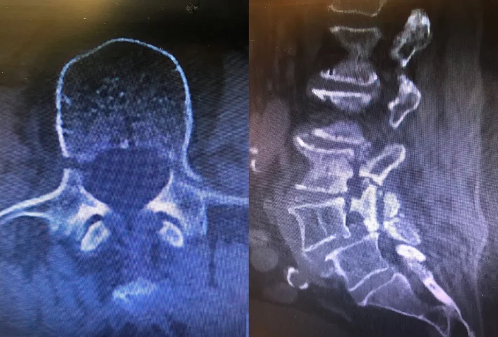
CT Scans
CT scans provide detailed images of the bone structures and are highly effective in detecting both early and advanced fractures. Fine-cut CT scans are particularly useful for assessing the severity and precise location of the fracture.⁸⁻⁹
MRI
MRI is excellent for visualizing soft tissues and can detect bone edema, which is indicative of stress reactions or early-stage fractures. It is also useful for assessing any associated soft tissue injuries.¹⁻⁷
Bone Scintigraphy (SPECT)
Bone Scintigraphy (SPECT) nuclear imaging technique is used to detect areas of increased metabolic activity, which may indicate a fracture. It is especially useful in cases where X-rays and CT scans do not provide conclusive results.⁸
Advanced Techniques
Intraoperative CT and navigation systems can be used during surgical procedures to ensure precise screw placement and to confirm the reduction of fractures.⁴
By combining these diagnostic tools, healthcare providers can accurately diagnose pedicle fractures and plan appropriate treatment strategies. Early detection and treatment are crucial to prevent the progression of the fracture and to ensure effective healing.
Fracture of Pedicle Prevention
Preventing pedicle fractures involves a multifaceted approach that focuses on maintaining spinal health, improving bone density, and minimizing stress on the vertebrae. Here are several key strategies:
Strengthening Core Muscles
Strong core muscles provide better support for the spine, reducing the likelihood of fractures. Engaging in regular exercises that target the back and abdominal muscles can enhance stability and support.¹⁻¹⁰
Maintaining Good Bone Health
Ensuring adequate intake of calcium and vitamin D, along with regular weight-bearing exercises, can help maintain strong bones. Addressing conditions like osteoporosis, which weakens bones, is also crucial in preventing fractures.¹⁻¹⁰
Proper Posture and Body Mechanics
Using correct posture and body mechanics during daily activities, especially when lifting heavy objects, can reduce stress on the spine. Ergonomic adjustments in the workplace and at home can also help minimize unnecessary strain on the back.¹⁰
Avoiding High-Risk Activities
Limiting activities that put excessive stress on the lower back, such as contact sports and high-impact exercises, can prevent stress fractures. If these activities are necessary, using protective gear and ensuring proper technique is important.¹⁰
Regular Check-ups and Early Detection
Regular medical check-ups can help detect early signs of bone density loss or spinal issues. Early intervention and management can prevent minor stress reactions from progressing to complete fractures.¹⁻¹⁰
Physical Therapy
Engaging in physical therapy can help improve spinal alignment, flexibility, and strength. This includes exercises designed to enhance core stability and correct postural imbalances, which can reduce the risk of fractures.¹⁰
By incorporating these strategies, individuals can reduce their risk of pedicle fractures and maintain overall spinal health.
Fracture of Pedicle Conclusion
Fracture of the pedicle, while not as commonly discussed as other spinal injuries, can have significant implications for one’s health and quality of life. Understanding the causes and recognizing the symptoms early are critical steps in managing this condition. Detecting pedicle fractures accurately requires a combination of clinical evaluation and advanced imaging techniques. Early diagnosis is key to preventing complications and ensuring appropriate treatment strategies are implemented promptly.
Preventive measures, including maintaining bone health through proper nutrition and exercise, can significantly reduce the risk of such fractures. By staying informed about the causes, symptoms, and preventive strategies, individuals can take proactive steps to protect their spinal health. Early detection and intervention remain the cornerstones of effectively managing pedicle fractures, underscoring the importance of awareness and timely medical consultation.
Disclaimer: The information provided in this blog is for educational purposes only and is not intended as a substitute for professional medical advice, diagnosis, or treatment. Always seek the advice of your physician or other qualified health provider with any questions you may have regarding a medical condition.
Fracture of Pedicle References
- The Back Pain Blog. (n.d.). Understanding lumbar pedicle stress reactions and other injuries. Retrieved from https://thebackpainblog.com/understanding-lumbar-pedicle-stress-reactions-and-other-injuries/
- Radiopaedia. (n.d.). Bilateral lumbar pedicle fracture. Retrieved from https://radiopaedia.org/cases/bilateral-lumbar-pedicle-fracture
- Radiopaedia. (n.d.). Spinal fractures. Retrieved from https://radiopaedia.org/articles/spinal-fractures
- The Journal of Neurosurgery: Spine. (2020). Lumbar pedicle fractures: a review of literature. Retrieved from https://thejns.org/spine/view/journals/j-neurosurg-spine/33/2/article-p199.xml
- American Academy of Orthopaedic Surgeons. (n.d.). OrthoInfo. Retrieved from https://orthoinfo.aaos.org/
- Spine Connection. (n.d.). Spinal & Artificial Disc Replacement Surgery. Retrieved from https://spineconnection.org/
- Hospital for Special Surgery. (n.d.). Spondylolysis (Pars Stress Fracture of the Spine). Retrieved from https://www.hss.edu/condition-list_spondylolysis-pars-fracture.asp
- Musculoskeletal Key. (n.d.). Stress fractures of the lumbar spine. Retrieved from https://musculoskeletalkey.com/stress-fractures-of-the-lumbar-spine/
- Röllinghoff, M., Zarghooni, K., Dargel, J., Hoffmann, M., Comstock, C., Burger, C., & Sobottke, R. (2008). Stress fractures of the lumbar spine. Archives of Orthopaedic and Trauma Surgery, 129(10), 1479-1485. https://doi.org/10.1007/s00402-008-0685-8
- Los Angeles Spine Institute. (n.d.). What are the treatments for lumbar stress fracture? Retrieved from https://www.laspine.com/what-are-the-treatments-for-lumbar-stress-fracture/