Spinal Navigation: A Comprehensive Guide for Improved Surgical Precision
Revolutionizing Surgical Precision in Spine Procedures with Spinal Navigation
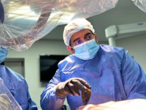
Spinal navigation has emerged as a groundbreaking technology that revolutionizes the field of spine surgery. With its ability to enhance surgical precision and accuracy, spinal navigation is transforming the way spine procedures are performed. In this comprehensive guide, we will explore the intricacies of spinal navigation, its mechanics, benefits, applications, limitations, and future directions, shedding light on the promising era of surgical precision.
What is Spinal Navigation?
Spinal Navigation Explained: Advancements in Precision Surgery
Spinal navigation is an advanced surgical technique that utilizes real-time imaging and tracking systems to assist surgeons in performing spine procedures with unparalleled precision. By providing detailed 3D visualization and navigation guidance, it allows surgeons to navigate the complex spinal anatomy with enhanced accuracy. Spinal navigation has significantly improved the safety and outcomes of various spine surgeries, including spinal fusion, tumor resection, and deformity correction.
How Does Spinal Navigation Work?
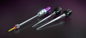
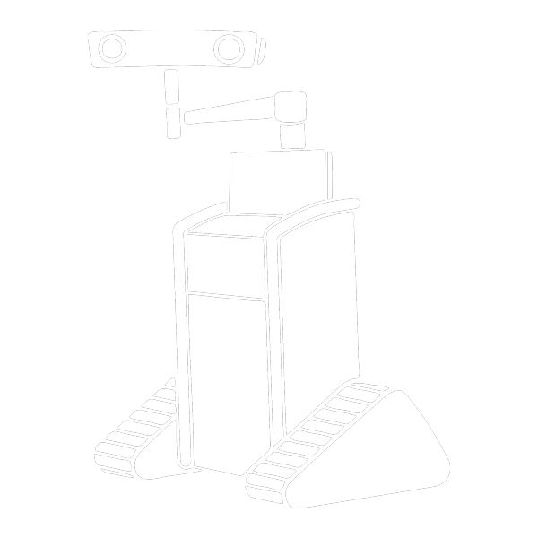
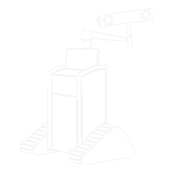
Spinal navigation systems employ a combination of imaging technologies, such as computed tomography (CT) scans and intra-operative fluoroscopy, along with specialized tracking devices. These devices are attached to surgical instruments and the patient’s anatomy, allowing real-time tracking and visualization of the surgical field. With the help of computer software, surgeons can precisely locate anatomical structures, navigate instruments, and ensure optimal implant placement, resulting in improved surgical outcomes.
Unlocking Surgical Precision: The Mechanics Behind Spinal Navigation
Spinal navigation encompasses various methods and techniques that aim to enhance surgical precision in spine procedures. These methods can be broadly categorized into two primary types: image-based navigation and electromagnetic navigation.
Image-Based Navigation
Image-based navigation relies on preoperative imaging, such as CT scans or MRI, to create a virtual 3D model of the patient’s spine. This model serves as a reference for the surgeon during the procedure, providing a roadmap for instrument placement and trajectory.
- CT-Based Navigation
This method utilizes CT scans to generate high-resolution 3D images of the patient’s spine. The navigation system then registers the virtual model with the patient’s actual anatomy to create an accurate representation. During surgery, the surgeon can visualize the position of instruments in relation to the virtual model, enabling precise guidance and alignment.
- MRI-Based Navigation
In cases where soft tissue visualization is crucial, MRI-based navigation is employed. This method utilizes MRI scans to create a 3D model that captures detailed anatomical structures, including nerves, discs, and ligaments. MRI-based navigation is particularly useful for procedures involving spinal tumors or conditions affecting the spinal cord.
Electromagnetic Navigation
Electromagnetic navigation utilizes electromagnetic fields to track the position and orientation of instruments in real-time during surgery. This method involves the use of electromagnetic sensors or coils, which are attached to the surgical instruments. The navigation system emits electromagnetic waves, and the sensors detect these signals, allowing precise tracking and visualization.
- Optical Navigation
Optical navigation systems use infrared cameras to track the movement of instruments equipped with reflective markers. These markers reflect infrared light emitted by the cameras, allowing the navigation system to calculate the instrument’s position and orientation. Optical navigation provides accurate and real-time tracking, enabling surgeons to navigate the spine with enhanced precision.
- Hybrid Navigation
Hybrid navigation combines the advantages of both image-based and electromagnetic navigation. This method utilizes preoperative imaging to create a virtual 3D model of the patient’s spine, which is then registered with the patient’s anatomy. During surgery, electromagnetic sensors or coils are attached to the instruments, providing real-time tracking and overlaying the instrument position onto the virtual model.
Each method of spinal navigation has its strengths and is selected based on the specific needs of the procedure and the surgeon’s preferences. These advanced technologies, whether image-based or electromagnetic, offer precise guidance, visualization, and tracking capabilities, enabling surgeons to perform spine procedures with unprecedented accuracy.
The continuous advancements in spinal navigation methods have led to the development of integrated systems that combine multiple techniques, further enhancing surgical precision. These integrated systems aim to provide comprehensive solutions by leveraging the strengths of different navigation methods, offering surgeons a versatile and adaptable approach to meet the challenges of complex spine surgeries.
Benefits of Spinal Navigation
Enhancing Surgical Accuracy: The Key Advantages of Spinal Navigation
Spinal navigation offers several compelling benefits for both patients and surgeons. Firstly, it enables surgeons to plan procedures more accurately by visualizing the patient’s unique anatomy in 3D. This enhanced visualization minimizes the risk of complications and reduces the need for exploratory surgery. Secondly, spinal navigation improves the accuracy of instrument placement, leading to better fusion rates and improved patient outcomes. Lastly, it can reduce radiation exposure during surgery, benefiting both patients and surgical staff.
Benefits and Strengths of the Spinal Navigation Methods
Here’s a comparison of the benefits and strengths of image-based navigation and electromagnetic navigation:
Image-Based Navigation
CT-Based Navigation Benefits & Strengths:
- High-resolution 3D imaging provides detailed visualization of bony structures, facilitating accurate instrument placement.
- Ideal for procedures involving complex spinal deformities, trauma, or spinal fusion.
- Offers precise guidance for pedicle screw insertion and alignment.
- Can assist in identifying anatomical variations, such as anomalous vertebral arteries.
- Excellent for visualizing bony structures and assessing anatomical relationships.
- Provides accurate navigation guidance in cases where soft tissue visibility is not critical.
- Well-established and widely used in spinal surgery with a proven track record of success.
MRI-Based Navigation Benefits & Strengths:
- Offers superior visualization of soft tissue structures, including nerves, discs, and ligaments.
- Particularly useful for procedures involving spinal tumors, spinal cord conditions, or cases requiring precise identification of neural structures.
- Helps in preoperative planning and intra-operative decision-making for tumor resection, decompression, and complex spinal procedures.
- Provides detailed visualization of soft tissue structures, enabling precise navigation and reducing the risk of nerve injury.
- Helps in identifying tumor boundaries and critical neural structures for safer surgical interventions.
- Can aid in preserving neurological function and optimizing surgical outcomes in complex cases.
Electromagnetic Navigation
Optical Navigation Benefits & Strengths:
- Real-time tracking and visualization of instrument position and orientation.
- Offers accurate navigation in complex anatomical scenarios, including deformities and cases with limited visibility.
- Helps in precise pedicle screw placement and alignment.
- Provides flexibility in instrument selection, as markers can be attached to a variety of instruments.
- Enables real-time guidance and feedback during surgery, enhancing surgical precision.
- Allows for efficient and reliable navigation even when direct line of sight is obstructed.
- Offers high accuracy and can be integrated with image-based navigation for enhanced guidance.
Hybrid Navigation Benefits & Strengths:
- Combines the strengths of both image-based and electromagnetic navigation techniques.
- Provides comprehensive guidance by overlaying real-time instrument tracking on preoperative imaging.
- Enables accurate navigation in complex cases, especially when both bony and soft tissue structures need to be visualized.
- Enhances surgical precision and reduces the risk of complications.
- Offers a versatile approach by leveraging the benefits of multiple navigation methods.
- Provides real-time feedback and guidance while incorporating the advantages of both image-based and electromagnetic navigation.
- Allows for precise navigation in challenging scenarios, leading to improved surgical outcomes.
It is important to note that the choice of navigation method depends on factors such as the surgeon’s expertise, the specific surgical procedure, patient characteristics, and available resources. Surgeons and healthcare professionals may select the most appropriate navigation method based on the specific needs of the surgery to achieve optimal outcomes.
Applications of Spinal Navigation
Spinal navigation has found applications in various spine procedures. It is commonly used in spinal fusion surgeries to guide the placement of screws, cages, and other implants. Additionally, it aids in the resection of spinal tumors and the correction of spinal deformities. Recent advancements in spinal navigation technology have expanded its utility in minimally invasive procedures, enabling surgeons to perform complex surgeries through smaller incisions, reducing tissue trauma, and enhancing patient recovery.
Preparing for Spinal Navigation
Optimizing Patient Outcomes: Preparing for Spinal Navigation Procedures
To ensure optimal outcomes with spinal navigation, thorough preoperative planning is essential. This includes obtaining high-quality imaging scans, such as CT or magnetic resonance imaging (MRI), to generate accurate 3D models of the patient’s spine. Additionally, meticulous registration of the imaging data with the patient’s anatomy and calibration of the tracking devices are crucial steps in preparing for spinal navigation procedures. Proper training of the surgical team is also imperative to maximize the benefits of this advanced technology.
Limitations and Challenges
While spinal navigation offers significant advantages, it is not without limitations. One of the challenges is the potential for registration errors, which may occur due to patient movement or anatomical deformities. Surgeons must be vigilant in verifying the accuracy of the navigation system throughout the procedure. Additionally, the cost of implementing and maintaining spinal navigation technology can be a barrier for some healthcare facilities. However, ongoing advancements in the field are addressing these challenges and making spinal navigation more accessible and reliable.
Future Directions and Innovations
The Future of Spinal Navigation: Cutting-Edge Technologies and Innovations
The future of spinal navigation holds great promise, with ongoing research and development focused on further improving surgical precision. One area of innovation is the integration of artificial intelligence (AI) algorithms into navigation systems, enabling real-time feedback and predictive modeling during surgery. Additionally, advancements in augmented reality and robotics are poised to enhance the capabilities of spinal navigation, allowing for even greater precision and less invasive procedures.
Conclusion
Transforming Spine Surgery: Unveiling the Promising Era of Spinal Navigation
Spinal navigation has ushered in a new era of surgical precision in spine procedures. By harnessing the power of real-time imaging, tracking systems, and computer-guided navigation, this technology has revolutionized the field of spine surgery. With its numerous benefits, including enhanced accuracy, improved patient outcomes, and reduced radiation exposure, spinal navigation has become an invaluable tool for surgeons. As we look ahead, ongoing advancements and innovations will continue to shape the future of spinal navigation, paving the way for even more remarkable achievements in the field of surgical precision.